
Scientific Activity (English)
Presently, the main research interest of Prof. Sergo is in the use of vibrational spectroscopies (Raman and IR) for the study of tissues and cells. This is a marked change from the past research activity which was mainly devoted to the study of advanced ceramic materials for structural and functional applications.
The main research areas are the following:
- Raman analysis of tissues with and without pathologies. The idea is to provide the anatomopathologist with a chemical information to be coupled with the histological sections. This activity is carried out with the fundamental collaboration of IRCSS Childrens Hospital Burlo-Garofolo;
- Raman SERS analysis of substances/drugs of interest in oncology; specifically we try to arrive at the determination in the range of nanomolar. This activity is carried out with the fundamental collaboration of CRO oncological hospital Aviano.
- Raman SERS study of quantum dots. In this activity we aim at making Raman SERS a viable tool for the analysis of solids.
- Use of optical spectroscopies for the determination of phases and stresses in ceramic materials;
In the past some efforts have been dedicated to:
- Point defect analysis of oxide ceramics for functional applications;
- Catalytic properties of oxide ceramics.
Hereafther, a brief image about all the topics above will be presented.
Raman analysis of tissues.
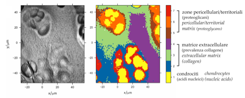
This figure reports the hystological section of a cartilage sample (left) and the result of a Cluster Analysis performed on about 5000 single Raman spectra acquired from the same section (right). The false color map has been obtained with the freeware HyperSpec, developed by Claudia Beleites while she worked in Prof. Sergo’s group (HyperSpec can be downloaded from hyperspec.r-forge.r-project.org). The picture has been taken from Analyst 135 (12), 3193-3204 (2010); DOI: 10.1039/C0AN00459F).
Raman SERS analysis of substances/drugs
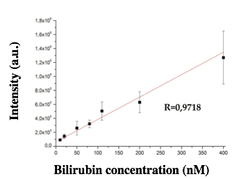
This figure reports the calibration curve for determining the concentration of free bilirubin from the intensity of the Raman SERS signal. This type of effort is under way for irinotecan, sunitinib and taxol, drugs widely used in oncology. The figure has been taken from Langmuir 28 (37), 13166-13171 (2012); DOI: 10.1021/la302383r).
Raman SERS study of quantum dots
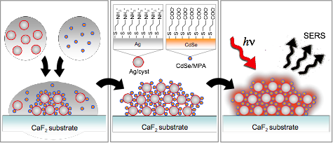
(Left) Formation of the electrostatically driven assembly of hybrid nanoparticles, obtained by mixing aqueous suspensions of cysteamine-capped Ag nanoparticles and MPA-capped CdSe Quantum Dots. (Center) Schematic of the hybrid nanoparticle assembly dried on the CaF2 substrate; the insets show the surface chemistry of the two components, leading to opposite electrical charge. (Right) SERS signal is obtained from the assembly. (Published on Journal of Nanoparticles Research vol 15 p. 1663 (2013); doi:10.1007/s11051-013-1663-9)
Use of optical spectroscopies for the determination of phases and stresses in ceramic materials
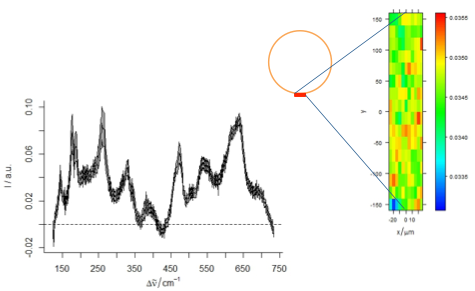
This is an example of the distribution of the monoclinic zirconia from an area of about 30 microns x 300 microns of a dental implant. Red spots are richer in monoclinic content, and the absolute values are in the range of 3-5% vol of monoclinic. The Raman spectra on the left report the mean and the Std. Dev. of the spectra (work performed within the LONGLIFE FP7 EU project).